
Figure 11.
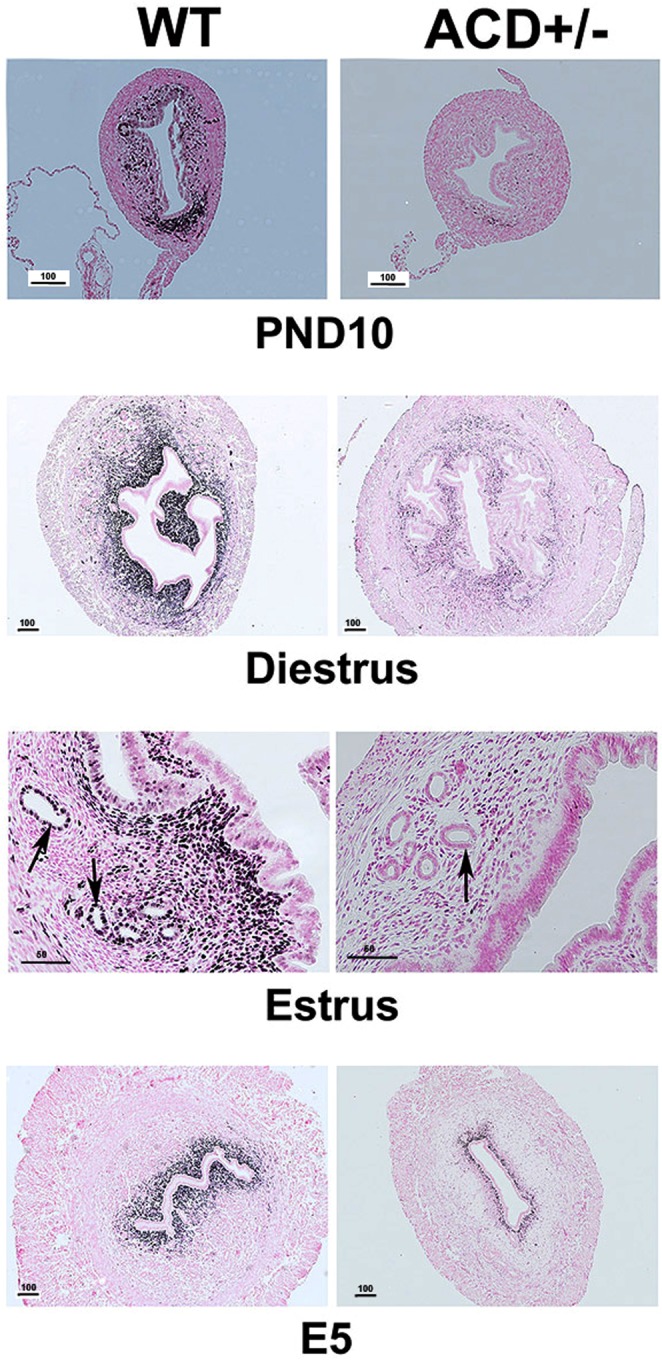
Immunohistochemical analysis of Lef1 expression. At PND10 there was robust Lef1 expression in uterine stromal cells, in particular at the mesometrial pole. In ACD+/− mutants (right panels) Lef1 expression was greatly reduced. Reduced Lef1 expression in mutants was also seen in adults at diestrus, estrus and at E5 post fertilization. During estrus there was Lef 1 expression in GE as well as stroma in WT uterus. Arrows point to uterine glands. Scale bars in microns.