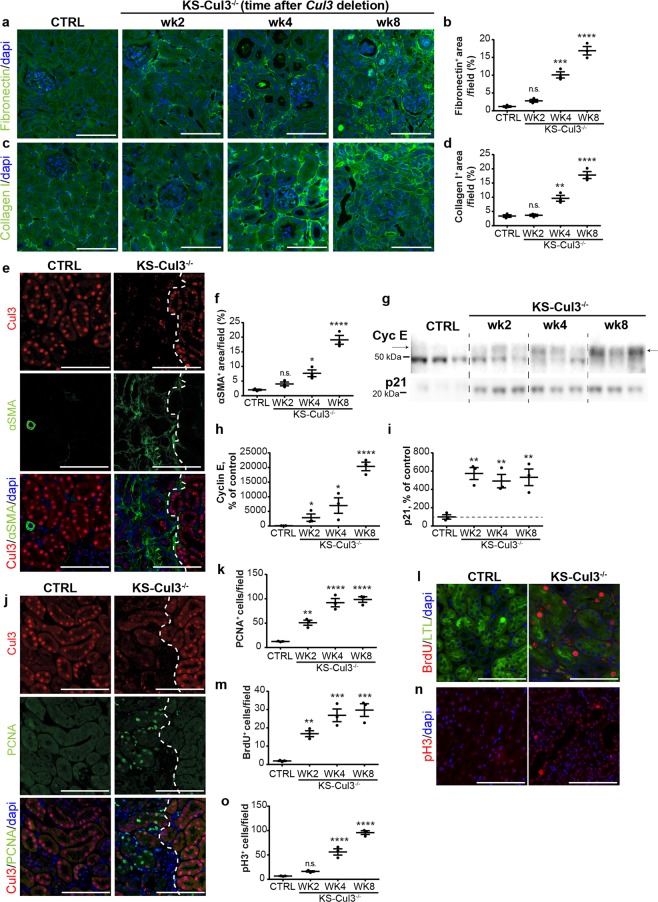
Figure 4.
Cul3 disruption causes progression into renal fibrosis and persistent cell cycle dysregulation. (a–d) Immunofluorescence staining revealed accumulation of fibronectin (a,b) and collagen I (c,d) in kidney-specific Cullin 3 knockout (KS-Cul3−/−) mice. (e,f) Cul3-deficient tubules were adjacent to areas of increased alpha smooth muscle actin (α-SMA)+ extracellular matrix accumulation. (g–i) Western blotting showed persistent increase of cyclin E and p21 abundance in KS-Cul3−/− mice up to at least 8 weeks. (j,k) Proliferating cell nuclear antigen (PCNA)+ cells, a marker for both DNA synthesis and DNA repair, were detectable within and nearby Cul3-deficient tubules. (l,m) Increased Bromodeoxyuridine (BrdU) incorporation indicated greater number of proliferating cells in S-phase upon Cul3 deletion in LTL+ proximal tubules. (n,o) Cul3 deletion was associated with increased number of tubule epithelial cells positive for phospho-Histone 3 (pH3), a marker for cells in G2 and M phase. Scale bars = 100 μm. Mean values are shown ± SEM. n = 3. Asterisks show significant differences between control and each KS-Cul3−/− mice group. *P ≤ 0.05, **P ≤ 0.01, ***P ≤ 0.001, ****P ≤ 0.0001; ordinary one-way ANOVA with Dunnett’s multiple comparisons test. Abbreviation: n.s., not significant. Coomassie-stained gels used for Western blot normalization can be found in Supplementary Fig. S4. Western blots were cropped for clarity; uncropped images can be found in Supplementary Fig. S7.

