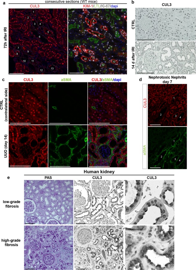
Figure 7.
CUL3 expression is dysregulated in AKI/CKD mouse models and human kidney tissue. (a) Immunofluorescence of consecutive kidney sections from control mice 72 h after IRI revealed decreased signal for CUL3 in most KIM-1+ tubules. n = 3. (b) Immunohistochemistry revealed strong CUL3 signal in sham mice in contrast to tubule cells 14 days after IRI. n = 3. (c) 14 days after unilateral ureteral obstruction (UUO), tubules had lower CUL3 signal and were adjacent to areas of increased alpha smooth muscle actin (α-SMA)+ extracellular matrix accumulation. n = 3. (d) Similar to (c), 7 days after injection of nephrotoxic serum, tubules had lower CUL3 signal and were adjacent to α-SMA+ areas. n = 3. (e) Human kidney tissue was obtained from tumour nephrectomy specimens and Periodic acid-Schiff stained sections of non-tumour renal tissue were assigned to the low-grade versus high-grade fibrosis groups. Immunohistochemistry revealed reduced CUL3 signal in high-grade fibrosis compared to low-grade fibrosis group. n = 3. Scale bars = 100 μm.

