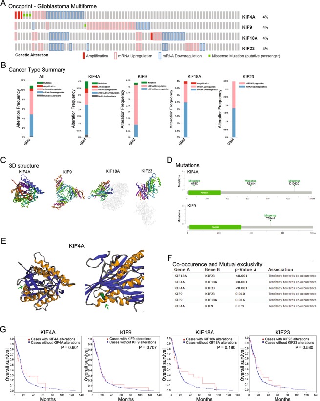
Figure 3.
Genomic analysis of KIF4A, 9, 18A, and 23 in GBM. The oncoprints of KIF4A, 9 18A, and 23 were identified. Genetic alteration of KIF4A, 9, 18A, and 23. Column represents GBM patients, and row represents gene alteration including amplification, deep deletion, mRNA upregulation, and missense mutation. (B) Genetic alteration summarized according to cancer type of GBM. (C) The 3D crystal structure of KIF4A, 9, 18A, and 23 is shown in rainbow colors (from N- to C- terminal ends). (D) The mutations of KIF4A and 9 were plotted. The kinesin motor domain is displayed in green with 15–336 amino acids. In the 75 amino acid sequence of KIF4A, Glycine was changed to Cysteine. Arginine, the 631th amino acids, was changed to Histidine in KIF4A. Aspartate, the 1062th amino acid, was changed to Glycine in KIF4A. In KIF9, Tyrosine, the 524th amino acid, was changed to Histidine. (E) The 3D structure of KIF4A implemented with missense mutations was plotted. In the kinesin motor domain of KIF4A, the mutated part was marked with a green arrow. (F) Co-occurrence and mutual exclusivity were examined for KIF4A, 9, 18A, and 23. (G) Kaplan–Meier survival analysis of KIF4A, 9, 18A, and 23 was conducted with and without alterations in overall survival. The p-values for Kaplan–Meier curves were calculated using the log-rank test.

