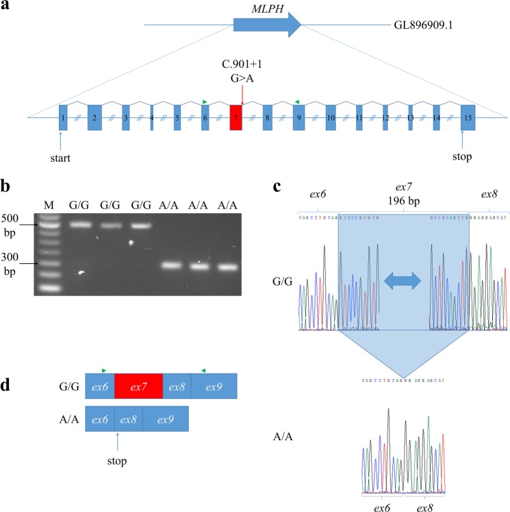
Figure 2.
Effects of MLPHp mutation on MLPH transcripts. (a) Structure of MLPH gene. Red box indicates exon 7. Green triangle indicates primers used for RT-PCR. Equal introns sizes are shown for simplification. (b) Agarose gel electrophoresis of MLPH cDNA exons 6–9. M – 50 bp DNA Ladder (NEB, USA). (c) An electrophoregram of Sanger sequencing for MLPH cDNA exons 6–9. Blue frame is exon 7 deleted in Silverblue (p/p) minks with homozygous MLPHp mutation. (d) Effects of MLPHp mutation on MLPH transcripts. Green triangle indicates primers used for RT-PCR.

