Figure 1.
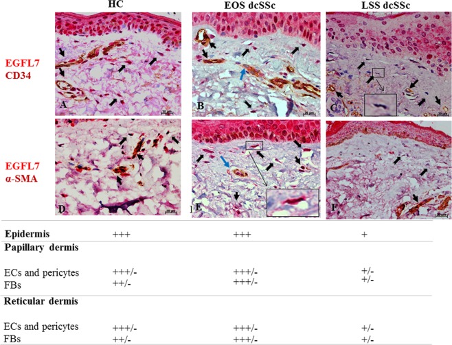
EGFL7 immunostaining in HC and dcSSc skin. (A,D) In HC skin, strong EGFL7 expression was detected in ECs and pericytes of vessels (black arrows), in fibroblasts of papillary and reticular dermis (black open arrows), and cells of the basal layer of the epidermis and keratinocytes. (B,E) In EOS dcSSc skin, strong EGFL7 expression was detected in ECs and pericytes of vessels (black arrows), in fibroblasts of papillary and reticular dermis and epidermal (black open arrows), cells of the basal layer of the epidermis and keratinocytes as in the HC skin. ECs of the most vessels were strong immunostained for EGFL7. However, ECs of a few vessels displayed a weaker immunostaining intensity for EGFL7 (blu arrows). Inset shows EGFL7 positive fibroblast (C and F) In LSS dcSSc skin, the remaining microvessels displayed a weak (or absent) immunostaining for EGFL7. Furthermore, fibroblasts of papillary and reticular dermis and cells of the basal layer of the epidermis and keratinocytes displayed a weak (or absent) immunostaining for EGFL7. Inset shows EGFL7 weak positive fibroblast. Semiquantitative scoring was performed independently by 2 blinded observers, based on the observation of immunostained skin sections. Scores are defined as follows: +++ intense staining; +++/− intense staining but not homogeneous positive staining; ++ moderate staining; + weak staining; +/− weak staining but not homogeneous positive staining.

