Figure 2.
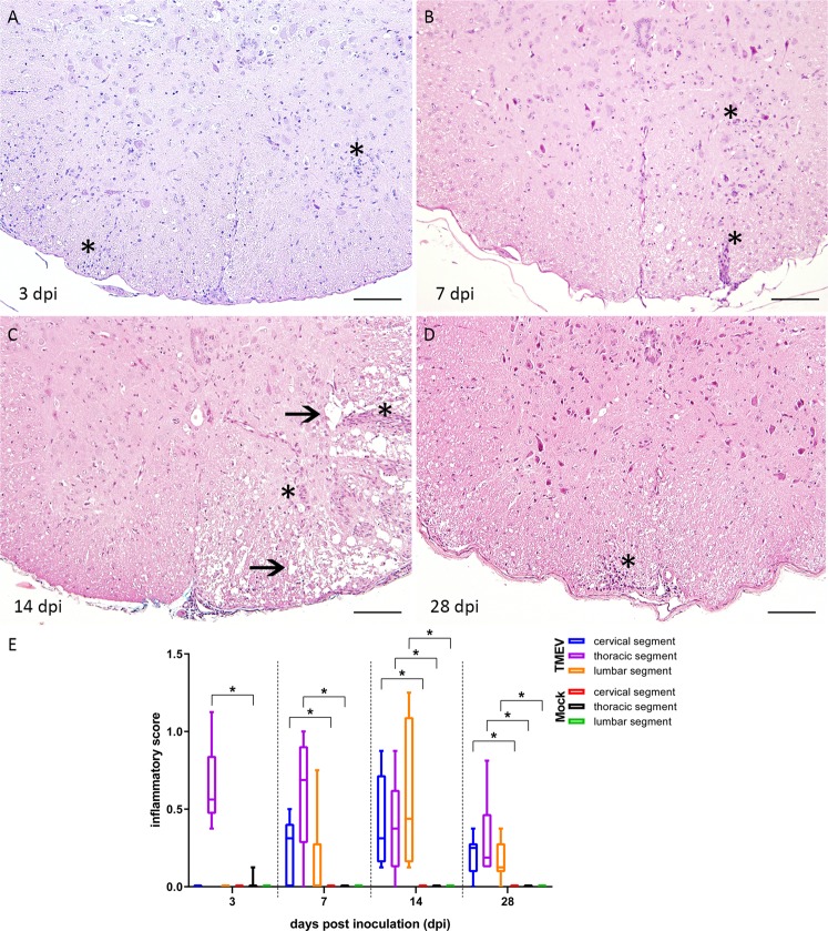
Histological changes within the thoracic spinal cord at 3 (A), 7 (B), 14 (C) and 28 (D) days post intraspinal infection with TMEV and the detection of an antero- and retrograde spread. (E) Lesions consisted of meningitis and perivascular accentuated lympho-histiocytic inflammation (A–D, asterisks) as well as multifocal dilated myelin sheaths and demyelination indicated by loss of eosinophilia in the white matter (C, arrows). The following number of animals was evaluated per group: n = 6–8 at 3 dpi; n = 5–6 at 7 dpi; n = 6–8 at 14 dpi; n = 6 at 28 dpi. Box-and-whisker plots show median and quartiles. Significant differences between the groups as detected by Mann-Whitney U-test are marked by asterisks (p ≤ 0.05). HE, bars = 100 µm.

