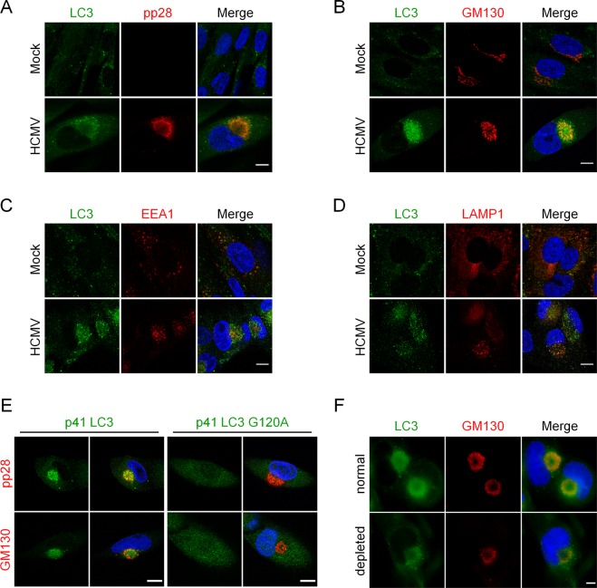
Figure 2.
Vesicles containing lipidated form of LC3 accumulate in the vAC. Confocal images of HFF mock-infected (Mock) or infected with HCMV AD169 strain at MOI 0.5 for 4 days and immunostained for LC3 and (A) pp28 (viral tegument protein), (B) GM130 (Golgi marker), (C) EEA1 (early endosome marker), or (D) LAMP1 (late endosome/lysosome marker). Representative images of 3 independent experiments. See also supplementary videos S2-4. (E) Confocal images of cells stably expressing p41-LC3 or p41-LC3 G120A infected with HCMV at MOI 0.5 for 4 days and immunostained for exogenous LC3 with pp28 or GM130. (F) Representative images of WT HFF infected with HCMV at MOI 0.5 for 4 days, and permeabilized either after fixation (“normal”) or before fixation (“depleted”) to eliminate soluble cytosolic proteins. Cells were immunostained for LC3 and GM130. Nuclei were subsequently stained with DAPI. Scale bar = 10 µm.

