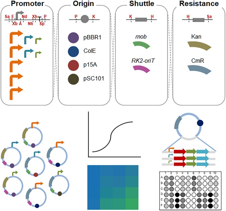
FIGURE 1.
Schematic diagram of the assembled shuttle vectors in S. oneidensis MR-1. The cutting sites of the restriction enzymes are shown in red. Sa, SacI; E, EcoRI; Xb, XbaI; A, AvrII; Nd, NdeI; Nh, NheI; Xh, XhoI; Sp, SpeI; P, PstI; K, KpnI; H, HindIII. High-strength promoters (i.e., pBAD, pXyl, pCI, pJ23119, and pTac) are shown in orange; medium-strength promoters (pTet and pTrc∗) are shown in aqua; and low-strength promoters (placUV5 and pLlacO1) are shown in olive.

