Figure 1.

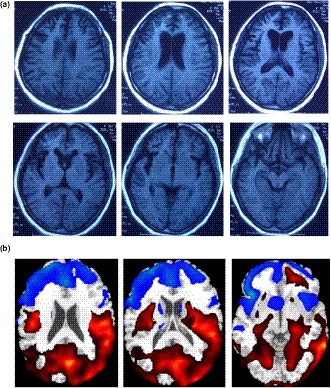
Imaging findings of the patient. (a) Cerebral MRI showed bilateral atrophy of frontal and temporal lobes. (b) FDG‐PET showed severe hypometabolism in frontal temporal areas

Imaging findings of the patient. (a) Cerebral MRI showed bilateral atrophy of frontal and temporal lobes. (b) FDG‐PET showed severe hypometabolism in frontal temporal areas