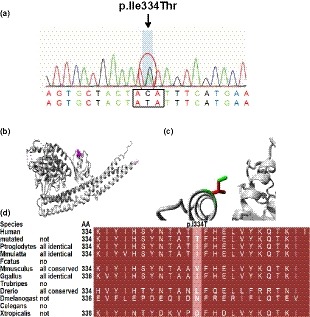
Figure 2.

Detection of p.Ile334Thr in TBK1 (NP_037386.1). (a) Sanger sequencing showed the heterozygous of the mutation. (b) The overview of protein. The protein is colored gray, and the side chain of the mutant residue is colored magenta. (c) Close‐up of the mutation. The protein is colored gray, and both the wild‐type (green) and mutant (red) residue are shown. (d) Conservation among multiple species at position 334
