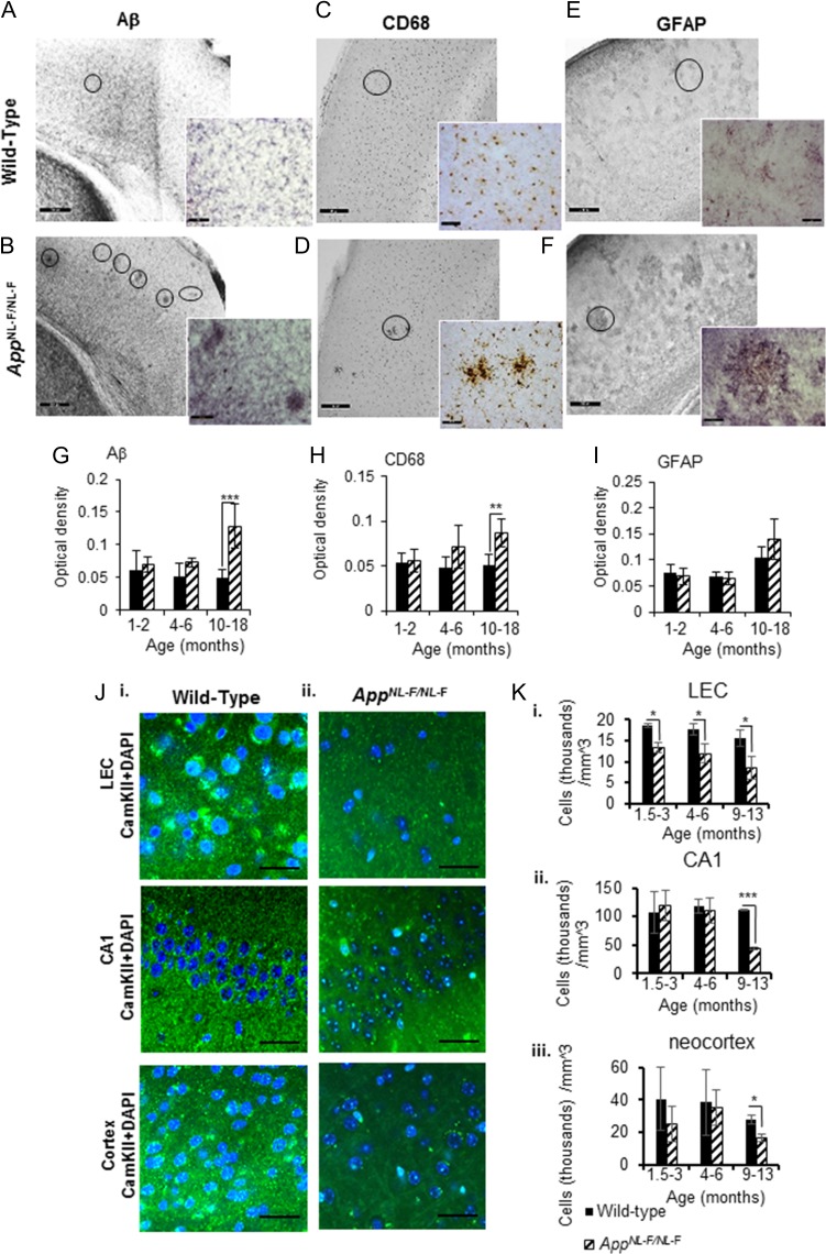
Figure 1.
The first knock-in AD mouse model, AppNL-F/NL-F shows an age-dependent accumulation of Aβ pathology, neuroinflammation and neurodegeneration of principal cells. (A) and (B) Immunoperoxidase labeling of Aβ in aged wild-type and AppNL-F/NL-F brains. Photographs taken with light microscope at 20× magnification; inserts illustrate photographs taken at 40× magnification. Circles show Aβ deposits. (C–F) Similarly, CD68 and GFAP labeling indicating microglia and reactive astrocytes respectively, in aged wild-type and AppNL-F/NL-F brains. The presence of strong Aβ aggregation in the AD mouse model, which correlates with increased inflammatory markers, is apparent (scale bar, 50 μm, enlarged images, scale bar, 20 μm). Circles indicate microglia and reactive astrocytes, respectively. (G–I) Graphs illustrating an age-dependent increase in Aβ, CD68, and GFAP levels in the 3 different age windows investigated in wild-type and AppNL-F/NL-F. AppNL-F/NL-F mice (10–18 months) demonstrated a significant increase in Aβ and CD68 level compared with wild-type aged animals, or the other 2 age cohorts (1–2 months, 4–6 months). Results are expressed as mean ± SD (**P < 0.01, ***P < 0.001; 2-tailed student t-test). (Ji-ii) Confocal imaging experiments using antibody to selectively label the expression of calcium/calmodulin-dependent protein kinase II (CamKII) expressed in principal excitatory cells in 3 cortical regions: LEC, CA1, and layer 2/3 of the neocortex. There is gradual decline in DAPI colocalised with CamKII (secondary antibody Alexa 488, green), suggesting neurodegeneration of principal cells in the AppNL-F/NL-F mouse model. Panels show representative confocal images taken at X63 magnification of tissue stained with CamKII and DAPI in wild-type animals at 13 months and in age-matched AppNL-F/NL-F mouse brains (scale bar = 20 mm). (Ki-iii) Pyramidal cell density was counted from collapsed confocal Z-stacks taken at ×20 magnification. There is significant reduction in pyramidal cell density in the lateral entorhinal cortex in the AppNL-F/NL-F animals compared with age-matched wild-type mice at all 3 ages investigated, which reduced significantly, (*P < 0.05, ***P < 0.001, 2-tailed Student t-test comparing AppNL-F/NL-F animals to wild-type counterparts separately at each of the 3 age groups investigated, n = 5 animals per cohort).

