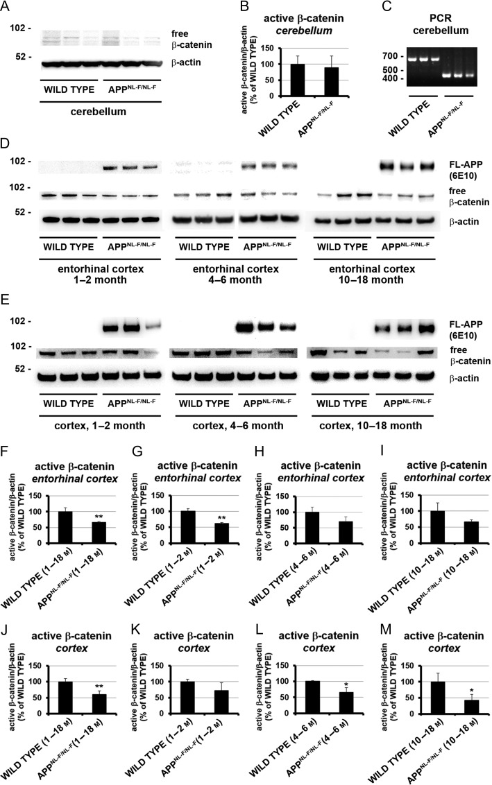
Figure 2.
Tissue and age-dependent Wnt signaling analysis of wild type and AppNL-F/NL-Fknock-in mice. Equal amounts of protein lysates from different brain regions were analyzed from 1 to 18 months old wild-type and AppNL-F/NL-F knock-in mice via immunoblotting. (A, D, and E) Representative Western blot analysis showing detection of active β-catenin in lysates from the cerebellum (A), entorhinal cortex (D), and cortex (E). Protein detection of human full-length APP (D and E) and mouse β-actin (A, D, and E) served as control. (C) Representative amplicon of 700 bp for wild type and 400 bp for AppNL-F/NL-F knock-in mice, confirming the genotype of animals used in (A) via polymerase chain reaction using genomic DNA. (B, F–M) Mean signal changes of active β-catenin in cerebellum (B), entorhinal cortex (F–I) and cortex (J–M). Western blot labeling showing age-dependent significant reduction of active β-catenin in the cortex of AppNL-F/NL-F knock-in mice compared with wild-type mice. Signals were divided through β-actin and normalized to wild-type (100%) represented as mean percentage ± SEM (*P < 0.05; **P < 0.01, n = 3–9).

