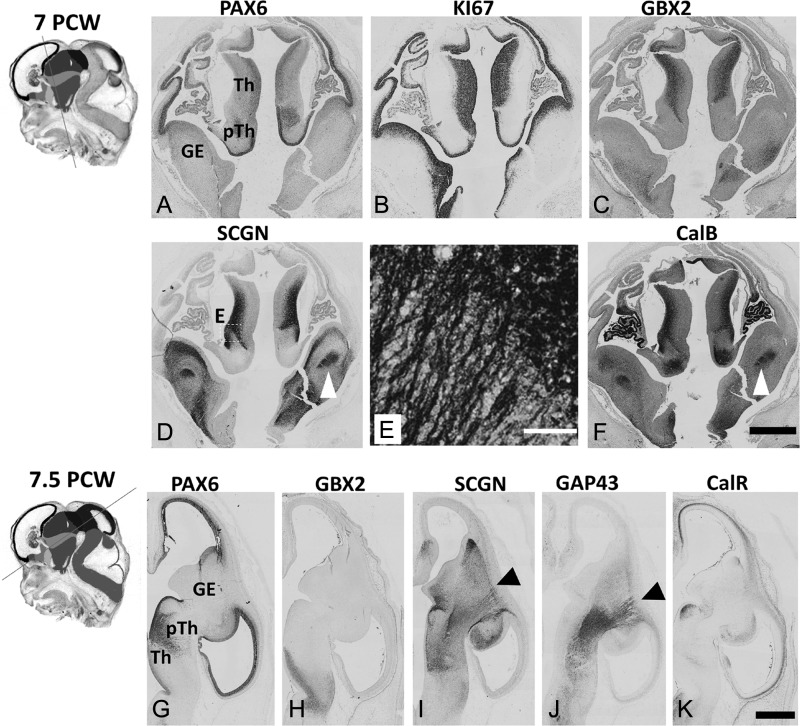
Figure 1.
Early development of the human thalamus. Insets show plane of sectioning (see Supplementary Fig. S1 for color version and key). (A–C). At 7 PCW in the thalamus (Th) PAX6 was moderately expressed in the VZ, KI67, a marker for dividing cells, was present in both VZ and SVZ whereas GBX2 was weakly expressed in the subventricular layer but strongly expressed in an outer post-mitotic mantle layer. The prethalamus (pTh) was characterized by strong expression of PAX6 in its ventricular zone (VZ). (D) SCGN was expressed in both cell bodies and neurites in the outer mantle layer of the thalamus, SCGN positive fibers also seen in the internal capsule (arrowhead). E is a higher magnifcation of the boxed area in (E). (F) similarly CalB was also expressed by post-mitotic thalamic neurons and in fibers running in the IC (arrowhead). By 7.5 PCW (G) PAX6 expression was maintained in thalamic VZ, while in the prethalamus PAX6+ cells were now seen away from the VZ forming a boundary with the thalamus. (H, I) GBX2 and SCGN immunoreactivity was present in post-mitotic cells of the thalamus which extend SCGN+ positive axons to the PSB (arrowhead). (J,L) These axons were also GAP43 positive, but there was very little expression of SCGN, CalB or GAP43 in the cortical IZ. (K) however CalR was expressed in the IZ, but this expression did not reach beyond the PSB. Scale bars: 1 mm in F (and for A–D); 100 μm in E; 1 mm in K (and for G–J).

