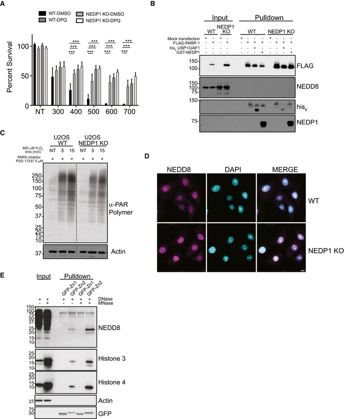
Figure EV4. PARP‐1 activity is reduced in NEDP1 KO cells which is protective against oxidative stress.

- PARP‐1 inhibitor DPQ (30 μM) protects WT cells from H2O2 treatment. NEDP1 deletion protects U2OS cells from H2O2 treatment greater than DPQ, and DPQ does not further protect NEDP1 cells from H2O2 treatment. Graphs represent the mean ± SEM of the percent survival compared to untreated cells. Two‐way ANOVA with Bonferroni post hoc test: n = 3, ***P < 0.0002.
- FLAG‐PARP‐1 is not modified by NEDD8. WT and NEDP1 KO U2OS cells were mock transfected or transfected with FLAG‐PARP‐1. After low induction with doxycycline for 24 h, cells were harvested and immunoprecipitation was performed with anti‐FLAG beads. After stringent washes, beads were split evenly and were treated with the deubiquitinase USP1/UAF1, the deneddylase GST‐NEDP1 or left untreated. Bound proteins were then processed for Western blot analysis with the indicated antibodies. There is some modified PARP‐1 that migrates as a higher molecular weight smear. The modification is reduced with deubiquitinase treatment but not with deneddylase treatment, which indicates that FLAG‐PARP‐1 is modified by ubiquitin. Furthermore, there is no immunoreactive of NEDD8 after immunoprecipitation, which indicates FLAG‐PARP‐1 is not modified by NEDD8 in either WT or NEDP1 KO cells.
- Reduction in PAR polymer accumulation in NEDP1 KO cells is not from increased PARG activity. U2OS WT and NEDP1 KO U2OS cells were pre‐treated for 1 h with the cell permeable PARG inhibitor PDD 17273 (5 μM) before treatment with H2O2 (600 μM). Cells were treated for the indicated amount of time, lysed and prepared for Western blot analysis with α‐PAR polymer antibody. PARG inhibition leads to increased PAR polymer accumulation in both cells lines as compared to cells without PARG inhibition (Fig 4A). However, the accumulation of PAR polymer in NEDP1 KO cells is still lower than in WT cells, which indicates PARP‐1 is not fully activated in NEDP1 KO cells.
- NEDD8 is predominantly nuclear in both WT and NEDP1 KO cells. U2OS WT and NEDP1 KO cells were plated on glass coverslips, fixed and prepared for immunofluorescence detection with α‐NEDD8 antibody and the DNA stain DAPI (scale bar = 10 μm).
- MNase digestion of DNA increases PARP‐1‐NEDD8 trimer binding. NEDP1 KO U2OS cells were harvested, and lysates were treated with DNase only or with DNase plus MNase. Treated lysates were then incubated with recombinant Zn1‐GFP or Zn2‐GFP for 1 h followed by immunoprecipitation with GFP‐Trap. Immunoprecipitated proteins were resolved by SDS–PAGE followed by Western blot analysis with the indicated antibodies.
