
Figure 5.
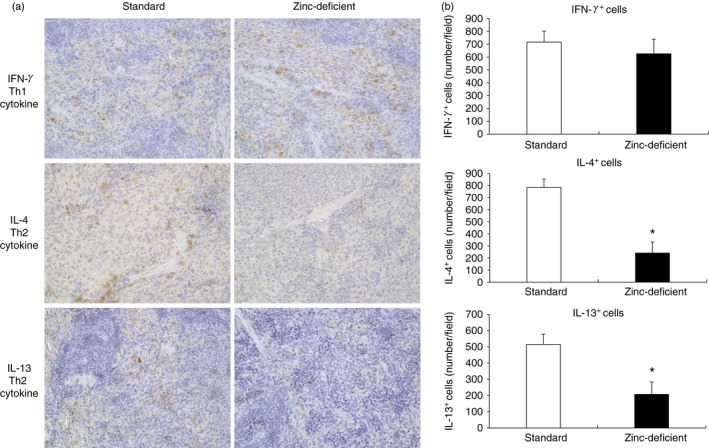
Immunohistochemistry for IL‐4, IL‐13, and IFN‐γ in spleens of rats on standard or zinc‐deficient diet. (a) Representative photomicrographs. Brown spots indicate immunohistochemical staining for IFN‐γ (a marker of Th1 cytokine), or IL‐4 and IL‐13 (markers of Th2 cytokines). Magnification, 400×. (b) Number of positive cells per field. Data represent the mean ± standard error (n = 5 per group). *P < 0·05 versus standard using the Mann–Whitney U‐test. IL, interleukin; IFN‐γ, interferon‐γ; Th1, T helper type 1.