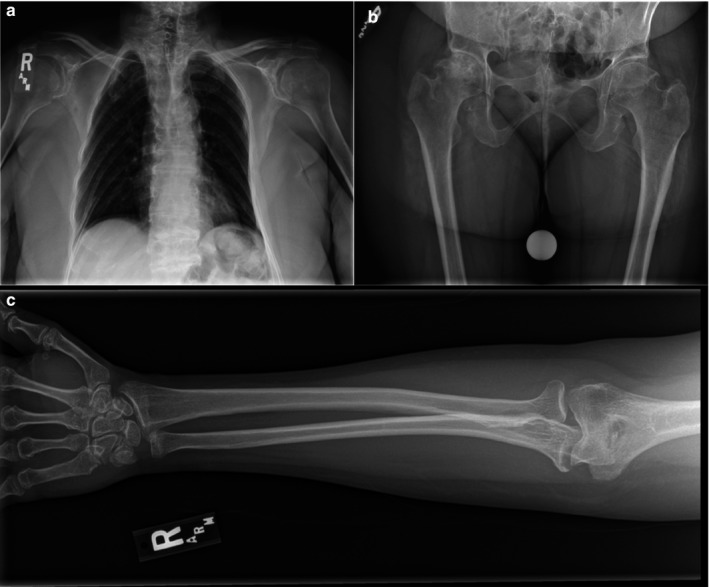
Figure 2.

X‐ray images of the proband. (a) Thoracic abnormalities as seen through X‐ray, note platyspondyly, asymmetry of ribs, and absence of scoliosis. (b) X‐ray of the hips, note reduced joint space between hip and right femur. (c) Right arm/hand of proband, metaphyseal abnormality is visualized
