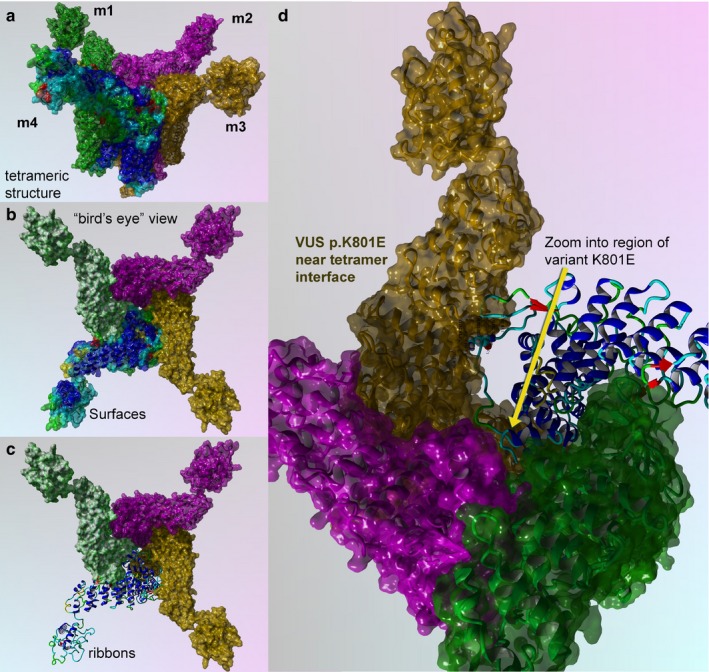
Figure 5.

TRPV4 surface and mapping for interaction for wild type and p.K801E variant. (a) Full‐length model for the entire TRPV4 tetrameric structure colored to distinguish the four protein monomer chains. Monomers 1–3 are colored uniformly, and monomer 4 is colored by secondary structure. (b) Bird's eye view for the wild‐type surface rendered model is shown. (c) Key region to zoom into is shown in ribbons and colored by secondary structure. (d) Zoom by 5X and rotation by the X‐Y plane at 135° are done to show position of the VUS (p.K801E), which has a critical point located near the center of the tetramer interface
