Figure 1.

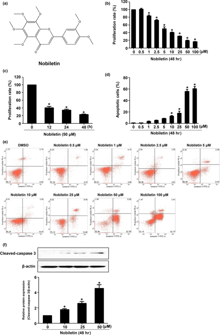
Nobiletin inhibited the cell viability and induced apoptosis in C666‐1 cells. The structure of nobiletin is shown in (A). C666‐1 cells were treated with gradient concentrations of nobiletin for 48 hr or with 50 μM nobiletin for indicated time periods. The cell proliferation rate was detected using CCK8 assay (B and C). The apoptosis rate was detected using flow cytometry (D and E). The expression of cleaved‐caspase 3 was measured with Western blot (F). *p < 0.05 vs. the group without treatment, n = 3
