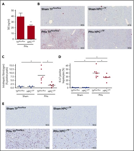
Figure 1.
Effect of liver TF deficiency on coagulation activation, fibrin(ogen) deposition, and liver regeneration after PHx. TFflox/flox mice and TFflox/flox/albumin Cre (HPCΔTF) mice were euthanized 30 minutes (n = 3 for sham, n = 6 for PHx) or 3 days (n = 6 for sham, n = 8 for PHx) after sham or PHx. (A) TAT plasma levels 30 minutes after PHx for TFflox/flox mice and HPCΔTF mice. (B) Representative images of fibrin(ogen) immunohistochemical staining 30 minutes after sham (upper panels) or PHx (lower panels). (C) Quantification of fibrin(ogen) deposition in TFflox/flox and HPCΔTF mice, expressed as percent positive pixel count. (D) Quantification of Ki-67–positive hepatocytes, expressed as the percentage of the total number of hepatocytes. (E) Representative images of Ki-67–stained livers for sham (upper panels) or PHx (lower panels) 3 days after surgery. Bars represent mean + SEM. Horizontal lines represent the mean, closed circles are individual mice. *P < .05.

