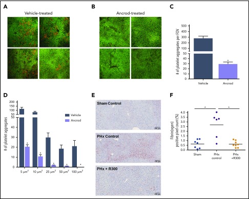
Figure 3.
Coagulation–platelet cross talk during liver regeneration after PHx. Platelets were visualized in the liver remnant using intravital microscopy. Platelets were labeled by IV injection of phycoerythrin-conjugated anti-mouse CD49b (red) immediately after PHx. Imaging was performed for 1 hour. Autofluorescent signal of the liver is shown in green to visualize liver anatomy. Representative images at 30 minutes after PHx in (A) vehicle-treated mice (n = 4) and (B) ancrod-treated mice (n = 4); images are from 4 individual mice per treatment group. Original magnification, 100×. Quantification of: (C) the number of platelet aggregates per field of view (FOV) and (D) the size equal to or larger than the indicated sizes. (E) Representative images of fibrin(ogen)-stained livers 30 minutes after sham (upper left panel) or PHx (upper right panel) for control mice, and 30 minutes after PHx for mice receiving R300 to deplete platelets (lower left panel). (F) Quantification of fibrin(ogen) deposition, expressed as percentage of positive pixel count, in livers of vehicle-treated mice (n = 7) or mice receiving R300 (n = 8) to deplete platelets. (F) Data are expressed as mean + SEM. *P < .05.

