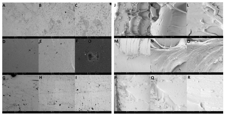
Figure 4.
(A–I) SEM images showing the surface morphology of (A–C) pure silk fibroin, (D–F) silk fibroin with 20% glycerol content, and (G–I) pristine PDMS films at 1000× (scale bar of 80 µm), 5000× (scale bar of 10 µm), and 10,000× (scale bar of 8 µm), respectively. (J–R) SEM images showing the cross-sections of (J–L) pure silk fibroin, (M–O) silk fibroin with 20% glycerol content, and (P–R) pristine PDMS at 1000× (scale bar of 80 µm), 5000× (scale bar of 10 µm), and 10,000× (scale bar of 8 µm), respectively.

