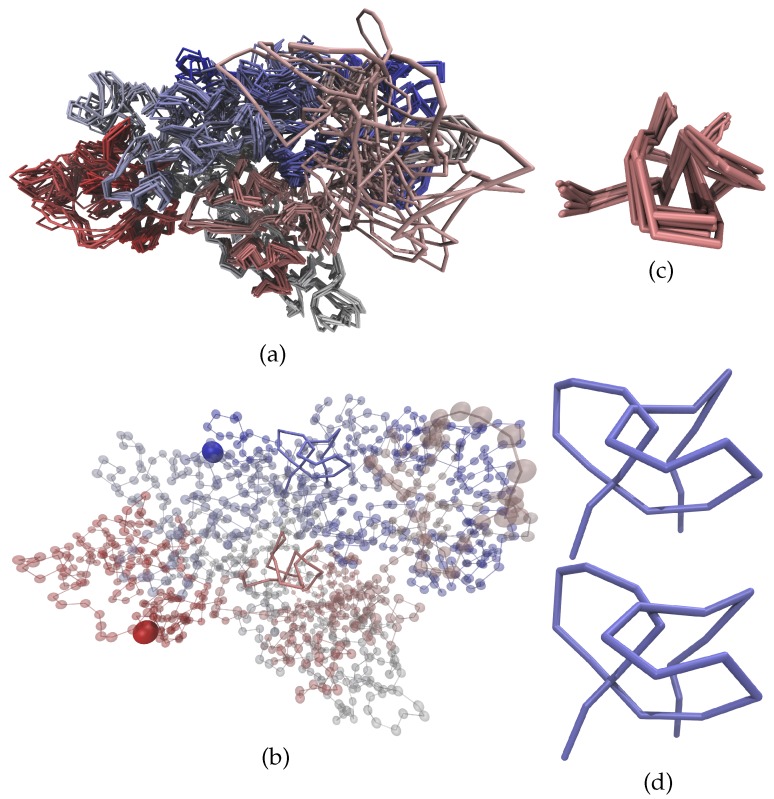
Figure 4.
Structure of chromosome 14 from cell 2 [48]. Panel (a) shows a comparison of all 10 models of the complete chromosome; whilst panel (b) shows one of those models (model 3), which contains two trefoil knots; Panel (c) shows an expanded view of one of the regions that forms a knot. Generally, the different models show a remarkable consistency. This corroborates our belief that the experimentally measured contacts define a non-degenerate ground state that is properly determined by the annealing procedure [48]. However, it is worth noting that there are also loops of the chromosome at the nuclear surface that do not exhibit many contacts. One example is the salmon colored loop in the top right of panel (a). Due to a lack of contacts this loop is not in a well-defined ground state but differs rather strongly between different models. In contrast, the two regions discussed in this paper do exhibit a well-defined structure. In panel (b), model 3 is represented as transparent beads with the knotted regions highlighted by solid lines—termini are accentuated by large solid beads. The red knot as shown in panel (c) has a very well-defined structure in all the models. In combination with the comparably large separations between the arcs of the knots, this leads to a consistent topology for all models. The structure of the blue region shown in panel (d) differs more strongly between the different models. In the upper center of the structure the separation between two of the arcs is very small. This leads to differences in the topology for different models as the trefoil knot that is present in some of the models (e.g., in the upper structure) can easily vanish (as shown in the lower structure) when the two arcs exhibit only a small relative change in their positions.

