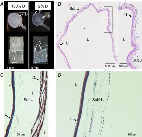
Figure 1.

The wall of murine detrusor‐free bladder preparation is intact and contains all layers except the detrusor smooth muscle and the serosa
A, photographs of intact and denuded bladder preparations filled with 200 μl KBS (upper panels) and dissected open at end of studies (bottom panels). B, a light microscopic view of hematoxylin and eosin‐stained denuded bladder wall demonstrating intact layers of the urothelium and submucosa. The area designated with black rectangle is magnified in the right panel. C and D, Masson's trichrome staining of filled intact (left) and denuded (right) bladders. The intact preparation has urothelium, suburothelium, detrusor smooth muscle and serosa whereas the denuded preparation has intact urothelium and suburothelium. The dashed line in D illustrates the boundary of the denuded preparation. D, detrusor smooth muscle; L, lumen; SubU, suburothelium; U, urothelium.
