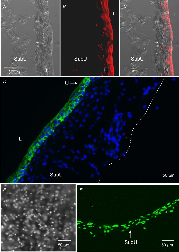
Figure 2.

The murine detrusor‐free bladder preparation is intact and contains specific cell types that are characteristic for urothelium and suburothelium
A–C, confocal microscopy of cryostat sections of the wall of a denuded bladder that was filled with sulfo‐NHS‐biotin (1 mg ml−1 PBS) for 30 min, then fixed in 4% paraformaldehyde and labelled with Alexa Fluor 594‐streptavidin (red, B and C). A is a differential interference contrast image of the same section, revealing no fluorescence in intermediate and basal cells and suburothelium. C is a merged image of A and B. Biotin–streptavidin fluorescence is limited to epithelia. Scale bar in A applies to A–C. D, cryostat section of denuded bladder wall showing cell nuclei (4′,6‐diamidino‐2‐phenylindole, blue) throughout the width of the preparation wall and localization of AQP3‐immunoreactivity (green) that is limited to basal cells and some intermediate cells of urothelium. Dashed line represents the outer edge of preparation. E and F, front wall (left) and cross section (right) of filled denuded bladder preparation from a PDGFRαEGFP mouse containing bright nuclei of eGFP‐tagged PDGFRα+ cells localized in suburothelium. L, lumen; SubU, suburothelium; U, urothelium.
