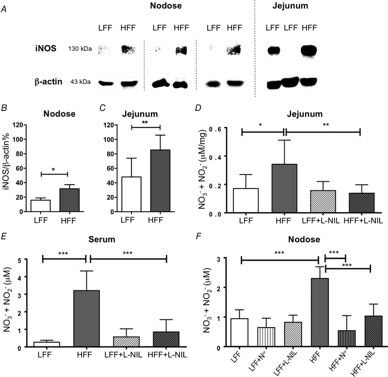
Figure 1.

iNOS expression and NO production were increased in HFF mice
A, total iNOS protein in nodose ganglion and jejunum visualized on SDS‐PAGE gel by western blotting. B and C, iNOS expression was increased in HFF mice nodose ganglia (B) (P < 0.05, n = 3 pools, each containing nodose ganglia from four mice, unpaired t test) and jejunum (C) (P < 0.01, N = 8). D and E, total nitrate/nitrite was increased in the jejunum (D) (P < 0.05, N ≥ 8, two‐way ANOVA with Bonferroni test) and serum (E) (P < 0.001, N = 6) from HFF mice. l‐NIL treatment (10 mg/kg, i.p. injection) reversed this change in both jejunum (D) (P < 0.01, N ≥ 7) and serum (E) (P < 0.001, N = 6). F, total nitrate/nitrite was significantly increased in culture media of nodose neurons from HFF mice compared to LFF mice (P < 0.001, N = 6, two‐way ANOVA with Bonferroni test). This augmentation in HFF nodose neurons was significantly attenuated by N ω (100 nm, P < 0.001, N = 6) and l‐NIL (10 μm, P < 0.001, N = 6).
