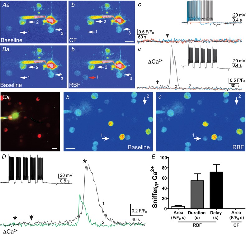
Figure 3.

Clustered but not continuous firing pattern evokes somatodendritic release of VP from individual neurones
A, pseudocolour image showing a patched eGFP–VP neurone surrounded by snifferVP cells (arrows, a, note the lack of snifferVP cell responses (b and c) when continuous firing (CF) activity was evoked in the patched neurone (arrowhead) using depolarizing pulses of increasing duration (c, inset). Ba and b, when the same neurone from A was then stimulated to evoke a repetitive bursting firing pattern (RBF, inset, Bc), a robust Ca2+ increase was observed in one of the snifferVP cells (Bc, Bb) approximately 30 s after stimulation (arrowhead). Ca, fluorescence image of a different patched eGFP–VP neurone loaded with Alexa 488 surrounded by snifferVP cells. Cb and c, corresponding pseudocolour images showing baseline (b) and snifferVP Ca2+ responses (c, arrows) when the patched neurone was stimulated to generate a RBF pattern (D, inset). D, plot of the snifferVP Ca2+ response over time to the RBF stimulation (arrowhead). Asterisks correspond to the time points of the images shown in Cb and c. E, summary data of the mean area, duration and delay of snifferVP responses to RBF stimulation (n = 31 snifferVP cells). Scale bars: 15 μm. [Color figure can be viewed at wileyonlinelibrary.com]
