Figure 4.

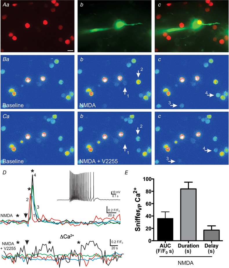
NMDA receptor activation evokes robust somatodendritic release of VP
A, fluorescence images of snifferVP cells (a) surrounding a patched eGFP–VP neurone loaded with Alexa 488 (b). Both images are superimposed in c. B, corresponding pseudocolour images showing snifferVP Ca2+ baseline (a) and responses in 4 cells (b and c, arrows) following focal application of NMDA (10 μm) to the patched neurone. Ca–c, NMDA stimulation to the same neurone failed to evoke snifferVP cell responses in the presence of the V1aR antagonist V2255 (1 μm). D, plots of snifferVP Ca2+ changes over time in the 4 cells shown in B and C, in the absence (upper panel) and presence (lower panel) of V2255. Arrowheads indicate the time of the NMDA stimulation, and asterisks correspond to the time points of the images shown in Ba–c and Ca–c. The inset shows the firing discharge of the patched neurone in response to NMDA stimulation. E, summary data of the mean area, duration and delay of snifferVP responses to NMDAR‐evoked firing in control ACSF (n = 21, from 10 patched eGFP–VP neurones). Scale bar: 15 μm. [Color figure can be viewed at wileyonlinelibrary.com]
