Figure 6.

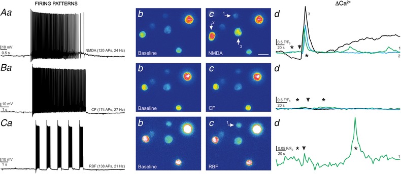
Somatodendritic release of VP is potentiated by NMDAR‐evoked firing when compared to the other firing modalities elicited in the same VP neuron
A, NMDAR‐evoked firing in a patched eGFP–VP neurone (NMDA 10 μm, a) resulted in positive Ca2+ responses in 3 snifferVP cells (b and c, arrows). The corresponding Ca2+ plots are shown in d. B, a continuous firing discharge (CF) evoked in the same eGFP–VP neurone (40 pA, single depolarizing pulse, a) failed to generate snifferVP responses (b–d). C, when the same eGFP–VP neurone was stimulated to evoke repetitive bursting firing (RBF, 80 pA, 5 depolarizing pulses, 0.5 s each, 1 s interval, a), a positive Ca2+ response was observed in only 1 snifferVP cell (b–d). The magnitude and delay of this response was evidently smaller and longer compared to the response observed following NMDAR‐evoked firing. Arrowheads in Ad, Bd and Cd show the time of the stimulation, and asterisks correspond to the time points of the images shown in panels b and c. [Color figure can be viewed at wileyonlinelibrary.com]
