Figure 7.

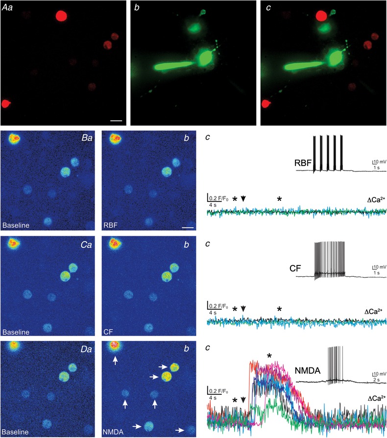
Representative example showing a more robust somatodendritic release of VP evoked by NMDAR‐mediated firing when compared to continuous or bursting firing in the same eGFP–VP neurone
A, fluorescence images of snifferVP cells (a) in the vicinity of a patched eGFP–VP neurone loaded with Alexa 488 (b). Both images are superimposed in c. B–D, corresponding pseudocolour images showing basal (a) and snifferVP Ca2+ responses when the patched neurone was stimulated to generate a RBF pattern (Bb), a CF pattern (Cb) or when firing was evoked by focal application of NMDA (10 μm, Db), in the sequence displayed. Plots of snifferVP Ca2+ responses to each of the stimulation protocols are shown in panels c. Note that in this eGFP–VP neurone, only NMDAR‐evoked firing resulted in robust Ca2+ responses in 7 different snifferVP cells (arrows, Db). Arrowheads in panels c indicate the stimulation time, and asterisks correspond to the time points of the images shown in panels a and b. Scale bar: 15 μm. [Color figure can be viewed at wileyonlinelibrary.com]
