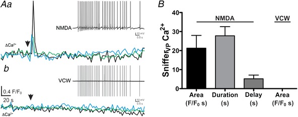
Figure 11.

NMDAR facilitation of somatodendritic release of VP is not dependent of the properties of the evoked somatic firing activity
Aa, plots of Ca2+ changes over time recorded from 3 snifferVP cells following focal application of NMDA (10 μm) to a recorded eGFP–VP neurone. The inset shows the firing response evoked in the patched neurone (31 APs, 3 Hz). Ab, the evoked activity in the patched neurone was then used as a voltage command waveform (VCW, inset) applied to the same neurone. The lower traces represent Ca2+ changes over time recorded from the same 3 snifferVP cells as in Aa following the VCW stimulation. Note the lack of snifferVP Ca2+ responses. B, summary data of the mean area, duration and delay in response to NMDA and VCW (n = 10 snifferVP cells from 5 patched eGFP–VP neurons). [Color figure can be viewed at wileyonlinelibrary.com]
