Figure 1.

TNX is expressed in neural structures in the stomach
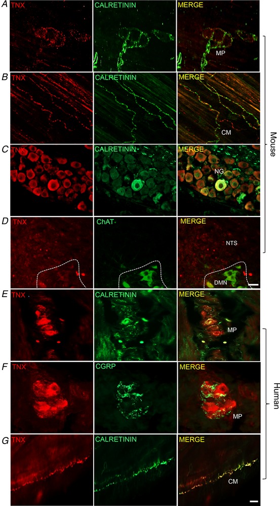
Representative immunohistochemical images taken from whole mount mouse fundus labelled with TNX and calretinin in IGLE surrounding the MP but not in cell bodies (A). TNX‐immunoreactivity was commonly found in calretinin‐immunoreactive fibres in the smooth muscle layer of the stomach (B, merge). The cell bodies of vagal afferents in the nodose ganglia also showed positive TNX and calretinin co‐labelling in sections (C, merge). TNX positive endings found in the mouse nuclus tractus solitarius (NTS) (D). Unlike mouse, human fundus sections showed TNX‐ and calretinin‐immunoreactive cell bodies in the myenteric plexus (MP) (E, merge) that were distinct from CGRP fibres (F, merge). TNX also co‐labelled calretinin‐positive endings in the circular smooth muscle (G, merge). All images from mouse and human stomach are whole mount confocal z‐images except for nodose ganglia (NG) and circular muscle (CM) images, which were taken with an epifluorescence microscope. DMN, dorsal motor nucleus. Scale bars in (A–D) = 25 μm, (E–G) = 30.
