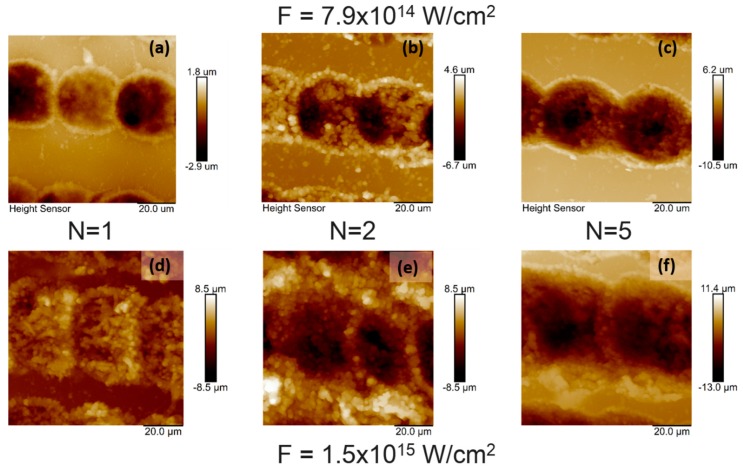
Figure 3.
Atomic force microscopy (AFM) topography images of chitosan thin film over an area of 20 × 20 µm2 irradiated with fs laser at λ = 800 nm, τ = 130 fs at different I and N: (a) I = 7.9 × 10¹⁴ W/cm2, N = 1, Rrms = 0.780 µm; (b) I= 7.9 × 10¹⁴ W/cm2, N = 2, Rrms = 2. 266 µm; (c) I = 7.9 × 10¹⁴ W/cm2, N = 5, Rrms = 2. 430 µm; (d) I = 1.5 × 10¹⁵ W/cm2, N = 1, Rrms = 1.617 µm; (e) I = 1.5 × 10¹⁵ W/cm2, N = 2, Rrms = 2.272 µm; (f) I = 1.5 × 10¹⁵ W/cm2, N = 5, Rrms = 2.480 µm.

