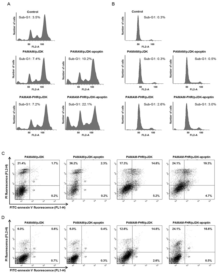
Figure 8.
Apoptosis of the PAMAM-FHR/pJDK-apoptin. (A,B) Cell cycle phase of PAMAM-FHR/pJDK-apoptin using propidium iodide (PI) staining. GBL-14 and dermal fibroblasts were treated with polymer/pJDK or pJDK-apoptin complexes for 36 h prior to flow cytometry. (C,D) Annexin V staining of PAMAM-FHR/pJDK-apoptin using FACS analysis. Each cell line was incubated under the same conditions as those used for cell cycle analysis. Q3 represents normal cells, Q4: early stage apoptosis, Q2: late stage apoptosis, and Q1: necrosis. PI, propidium iodide.

