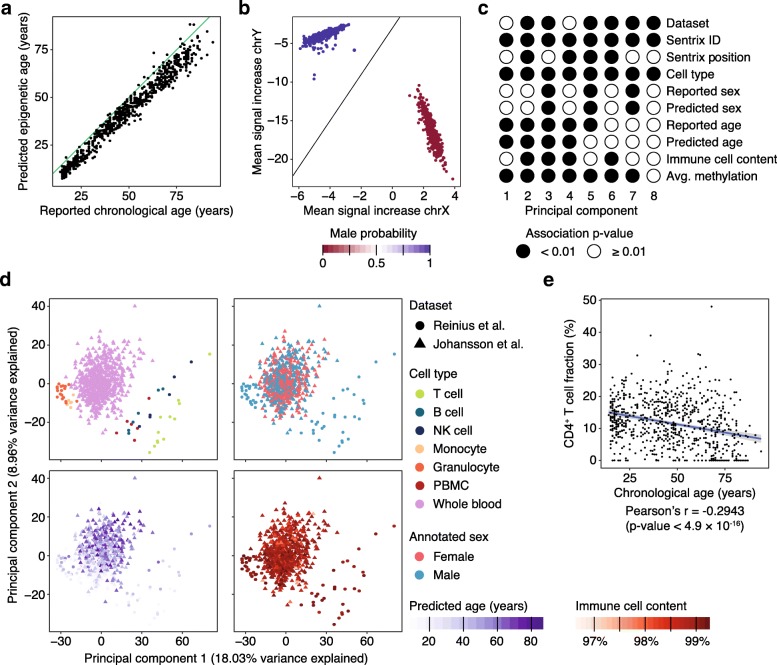
Fig. 2.
Analysis of a large DNA methylation dataset of blood samples profiled using Infinium 450k. a Scatterplot showing the correlation between epigenetic age predicted from DNA methylation and reported chronological age for 729 healthy donors (three individuals were excluded because no chronological age was reported). b Positioning of the samples in two-dimensional space for sex prediction. c Statistical association between principal components (columns) and sample annotations (rows). Significant associations with p values below 0.01 are marked by filled circles, while non-significant values are represented as empty circles. d Principal component analysis for 792 blood-based DNA methylation profiles, comprising 732 peripheral blood samples and 60 sorted blood cell populations, using the same principal components as in panel c. Immune cell content was estimated using the LUMP algorithm. e Scatterplot showing the negative correlation between chronological age and the estimated fraction of CD4+ T cells

