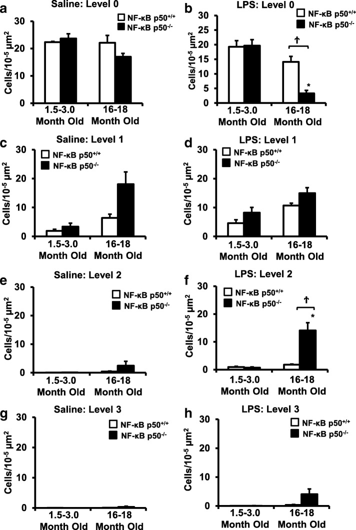
Fig. 4.
Aging NF-κB p50−/− mice have enhanced activated microglia morphology in response to peripheral LPS injection. Young adult (1.5–3.0 month old) and middle-aged (16.0–18.0 month old) NF-κB p50+/+ and NF-κB p50−/− mice were injected with saline or LPS (5 mg/kg, IP) to evaluate loss of NF-κB p50 function on microglia morphology in vivo. IBA1 stained microglia within the substantia nigra pars compacta (in the midbrain) were categorized into stages of activation ranging from resting (stage 0) to highly activated (stage 3). The relative number of microglia at 3 h post-injection within stage 0 (a, b), stage 1(c, d), stage 2(e, f), and g & h stage 3(g, h) was quantified by the fractionator method. Values are reported as mean cells/μm2 ± SEM of 3 coronal sections (40 μm) per animal. An asterisk indicates significant difference (P < 0.05) from control and a dagger indicates a difference between mouse strains. n = 3

