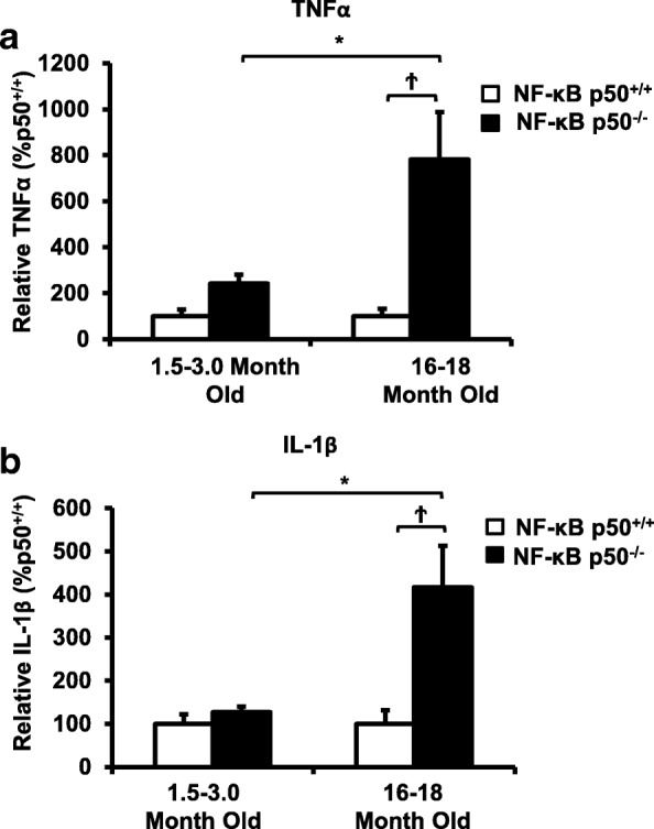
Fig. 6.

Loss of NF-κB p50 function and aging synergistically interact to amplify TNFα and IL-1β in isolated microglia. Young adult (1.5–3.0 month old) and middle-aged (16.0–18.0 month old) NF-κB p50+/+ and NF-κB p50−/− mice were injected with LPS (5 mg/kg, IP), and isolation of microglia from the whole brain with CD11b microbeads was performed at 3 h post-injection. Differences in a TNFα and b IL-1β were evaluated by quantitative RT-PCR. Values are normalized to GAPDH using the 2−ΔΔCT method and are reported as mean percent of NF-κB p50+/+ young adult saline control ± SEM. An asterisk indicates significant difference (P < 0.05) from the 1.5–3-month group and a dagger indicates a difference between mouse strains. n = 3
