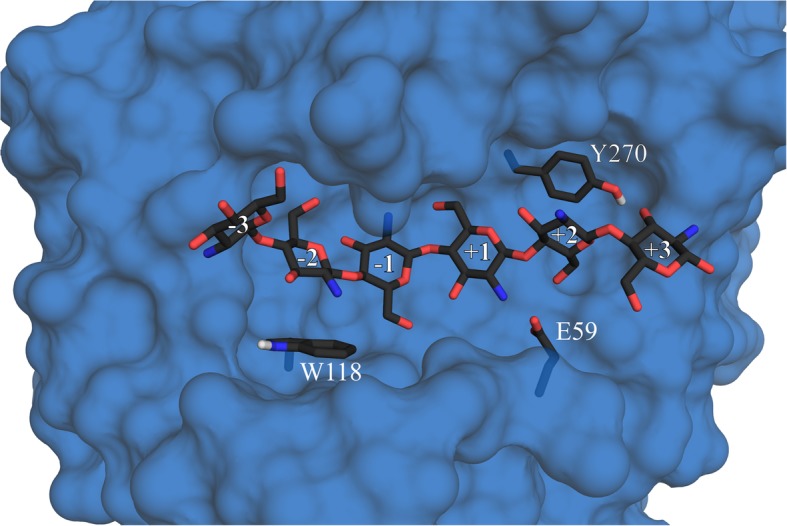
Fig. 1.

Selected CSN residues with a putative function in substrate binding. A three-dimensional homology-based model of CSN based on the crystal structure of Bacillus sp. K17 chitosanase (PDB: 1V5C, amino acid sequence identity: 97.47%), illustrating the side chain positions of the amino acids W118, E59, and Y270 relative to the docked substrate D6. The subsites (− 3) to (+ 3) are indicated. The enzyme surface without the side chains of the listed amino acids is pictured in blue, the labeled amino acid side chains emerging from the surface and D6 are colored by element.
