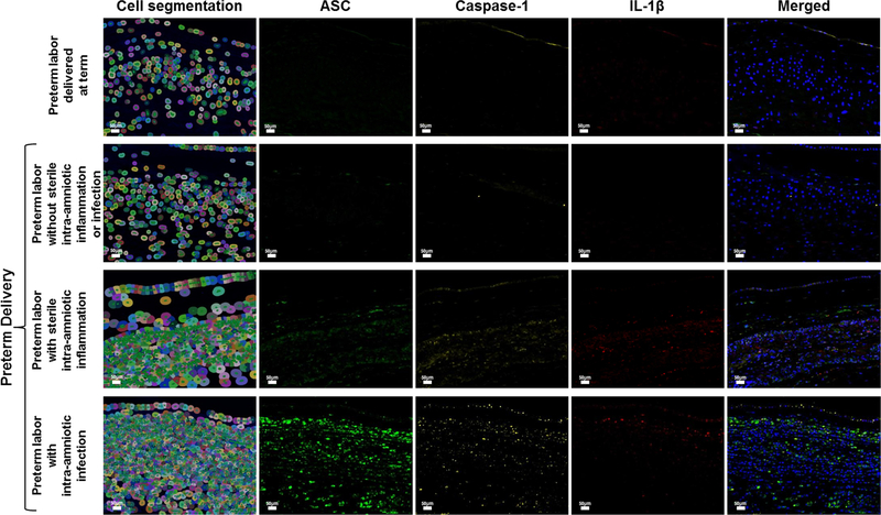
Figure 4.
Expression and co-localization of inflammasome components in the chorioamniotic membranes of women who underwent spontaneous preterm labor. Multiplex immunofluorescence staining of ASC (green), caspase-1 (yellow), and IL-1β (red) was performed in the chorioamniotic membranes of women who underwent spontaneous preterm labor but delivered at term and those who delivered preterm without sterile intra-amniotic inflammation or intra-amniotic infection, with sterile intra-amniotic inflammation alone, or with intra-amniotic infection. Phenoptics was performed to generate cell segmentation images as well as separate and merged immunofluorescence images. Images are representative of 3 experiments per group. Images were taken at 400X magnification and scale bars represent 50 μm.

