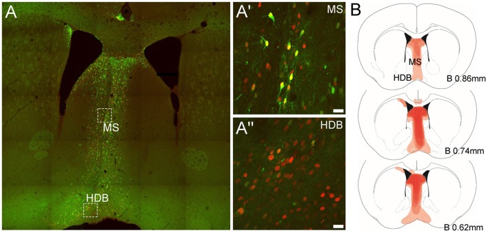
Figure 1.
Representative expression of green fluorescent protein (eGFP) and Cre-recombinase and viral spread in medial septum/diagonal band of Broca (MS/DB) in mice injected with the AAV(1/2)-Cre-IRES-eGFP viral vector. (A) Tiled (20× mosaic) image illustrating the presence of eGFP (green) and Cre-recombinase (red) in neurons in the MS/DB. (A′,A″) High magnification images (63× maximum projections) of boxed areas in (A) in MS and HDB, revealing the presence of endogenous eGFP and Cre-recombinase IR in both regions, with some neurons (yellow) strongly expressing both proteins and other neuronal cell-bodies expressing stronger Cre-IR than eGFP, consistent with the design of the viral vector used. (B) Schematic images documenting the relative level of Cre-recombinase expression (light/dark red shading) observed at different bregma levels containing the MS, HDB and VDB in the C57BL/6J floxed-Rxfp3 mice (n = 8) analyzed in the behavioral studies. Darker areas represent those sites where the viral-driven Cre-recombinase expression was consistently concentrated in most or all mice at that level.

