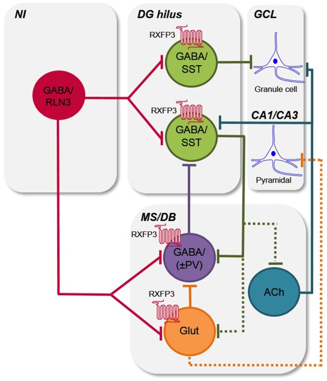
Figure 6.
Putative circuits by which relaxin-3/RXFP3 signaling modulates activity in the hippocampus and medial septum/diagonal band of Broca. Simple schematic circuit diagram to illustrate how the relaxin-3/RXFP3 system is anatomically positioned to modulate the activity of the septohippocampal and hippocamposeptal systems, which are important for cognitive functions and related behaviors (Tsanov, 2015; Korotkova et al., 2018; Müller and Remy, 2018). Based on previous and current findings (see “Discussion” section), the ascending relaxin-3 projections from the NI innervate local and/or projection GABA/SST neurons in the mouse DG hilus (Haidar et al., 2017), and the GABA/PV+ve hippocampal projecting neurons and possibly other GABA interneurons (i.e., GABA/(PV−ve), as well as some glutamate neurons in the MS/DB, and modulate their activity via RXFP3 (current study; see e.g., Müller and Remy, 2018 for recent review). Existing data also indicates that neurons present in the other nodes of the septohippocampal system (SHS) are RXFP3-positive, but as their neurochemical identity is currently unknown, they are omitted for clarity. Furthermore, any possible collateralization of relaxin-3 neurons and all possible intra-MS/DB interconnections are not illustrated for clarity. Abbreviations: ACh, acetylcholine; CA1/CA3, CA1 and CA3 layers of hippocampus; DG hilus, dentate gyrus hilus; GABA, γ-aminobutyric acid; GCL, granule cell layer; Glut, glutamate; MS/DB, medial septum/diagonal band of Broca; NI, nucleus incertus; PV, parvalbumin; RLN3, relaxin-3; RXFP3, relaxin-family peptide receptor 3; SST, somatostatin; ±, plus or minus (PV).

