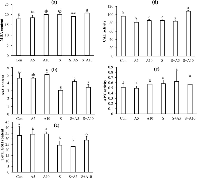
Fig. 4.
MDA content, nmol g−1 FW (a); AsA content, µmol g−1 FW (b); GSH content, µmol g−1 FW (c); CAT activity (d); APX activity (e) after 2 days of different treatments. Treatments were Control (Con), 5 mM Na-acetate (A5), 10 mM Na-acetate (A10), 100 mM NaCl (S), 100 mM NaCl+5 mM Na-acetate (S+A5), and 100 mM NaCl+10 mM Na-acetate (S+A10). Mean (± SD) were calculated from three replicates for each treatment. Values with different letters are significantly different at P ≤ 0.05 applying Fisher’s LSD test

