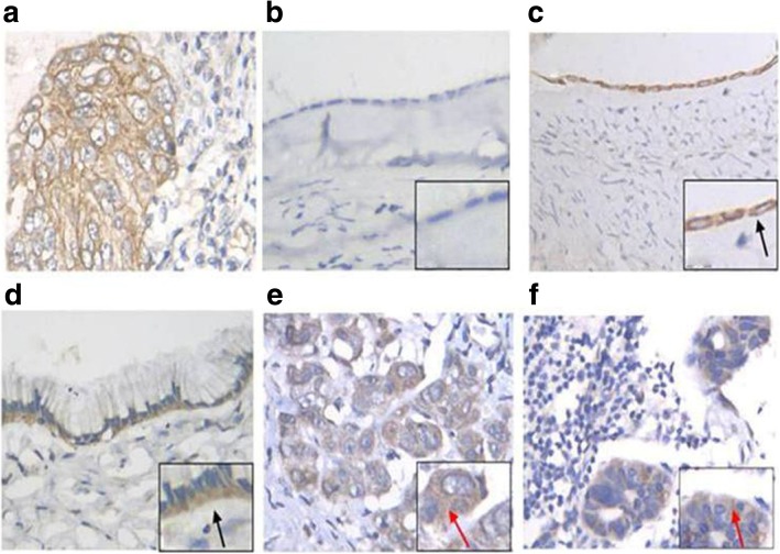
Fig. 1.
TM4FS1 expression in ovarian tissues and metastatic lymph node foci detected by IHC (400X). a Positive control. b Negative expression of normal ovarian epithelial tissues. c Positive expression of normal ovarian epithelial tissues. d Positive expression of ovarian benign tumor tissues. e Positive expression of epithelial ovarian cancer tissues. f Positive expression of metastatic lymph node foci. Black arrows: Expression of TM4FS1 is concentrated in the membrane or near the basement membrane of the cell membrane; Red arrows: Expression of TM4FS1 is concentrated in the cytoplasm

