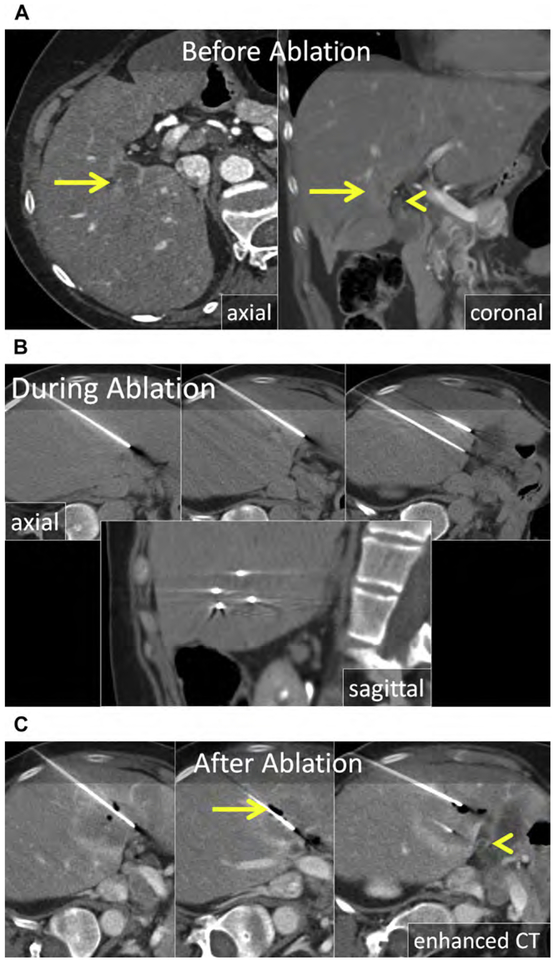
Fig. 10.
(A) Axial and coronal enhanced CT shows a CRLM in the medial right liver (arrow) in proximity to the common duct (arrowhead). (B) Axial and sagittal unenhanced CT following CT-guided placement of IRE electrodes. The electrodes are nearly parallel with near-equal spacing of 1 to 1.5 cm. Because precise electrode placement is important and a large number of electrodes are generally required for IRE, applicator placement can be time consuming. (C) Enhanced CT immediately following IRE. The ablation encompasses the tumor including a 10-mm margin and there is no apparent damage to the bile duct (arrowhead). The gas within the ablation zone (arrow) suggests that both thermal and nonthermal mechanisms of cell death were present in this case.

