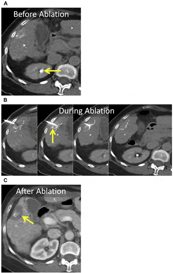
Fig. 13.
HCC in a patient with cirrhosis before, during, and after radiofrequency (RF) ablation. (A, B) Enhanced CTobtained for ablation planning immediately before RF electrode placement. Note that the contrast has washed out of the liver and is being excreted by the kidneys (arrow). (B) The only useful guide for placement of the RF electrode was retained ethiodized oil (arrow).(C)Enhanced CT1 month after RF ablation.The ablation encompasses the index lesion containing ethiodized oil, including a 5-mm margin (arrow).

