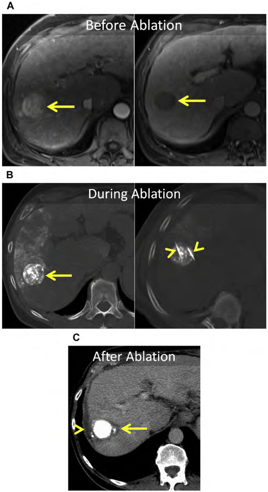
Fig. 15.
(A) Large (>3 cm) HCC in the right liver (arrow). (B) Unenhanced CT before and after placement of MW antennas (arrowheads). Note the excellent accumulation of ethiodized oil within the tumor (arrow) with partial clearance of ethiodized oil from the nontargeted liver in the 2-week interval between TACE and MW ablation. (C) Enhanced CT immediately following MW ablation. The ablation encompasses the tumor including a 5-mm margin (arrow). Artificial ascites was infused before and during MW ablation (arrowhead).

