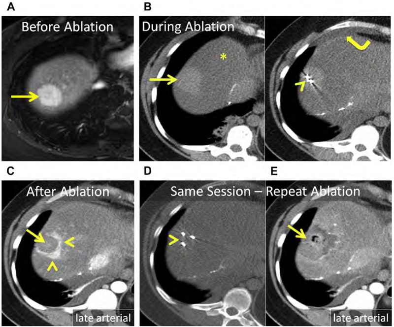
Fig. 18.
(A) Hepatic adenoma in the dome of the liver, Couinaud segment VIII (arrow). (B) Unenhanced CT after placement of 2 MW antennas (arrowhead). The mass (arrow) is conspicuous at unenhanced CT because of severe, diffuse hepatic steatosis (asterisk). Note artificial ascites used to protect the body wall and diaphragm (curved arrow). (C) Enhanced CT immediately following MW ablation. Persistent eccentric avid enhancement (arrowheads) surrounding the nonenhancing ablation (arrow) is compatible with viable tumor. (D) CT image following reinsertion of the 2 antennas and the addition of a third MW antenna (arrowhead). (E) Enhanced CT immediately following same-session repeat MW ablation. The ablation now encompasses the index lesion (arrow).

