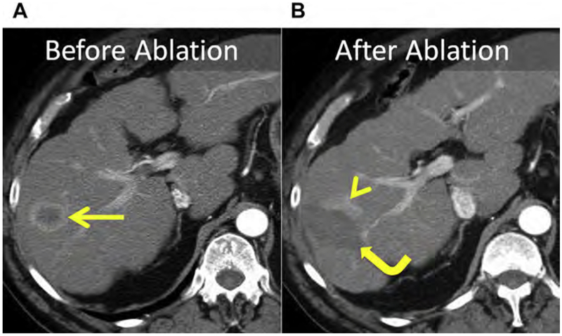
Fig. 19.
(A) CT before ablation shows an HCC within the right liver (arrow). (B) At follow-up, eccentric enhancement (arrowhead) adjacent to the index ablation (curved arrow) is consistent with LTP. Satellite tumors invisible at conventional imaging are generally the cause for LTP, suggesting that an adequate margin was not achieved. For HCC, a minimum of a 5-mm circumferential margin is necessary.

