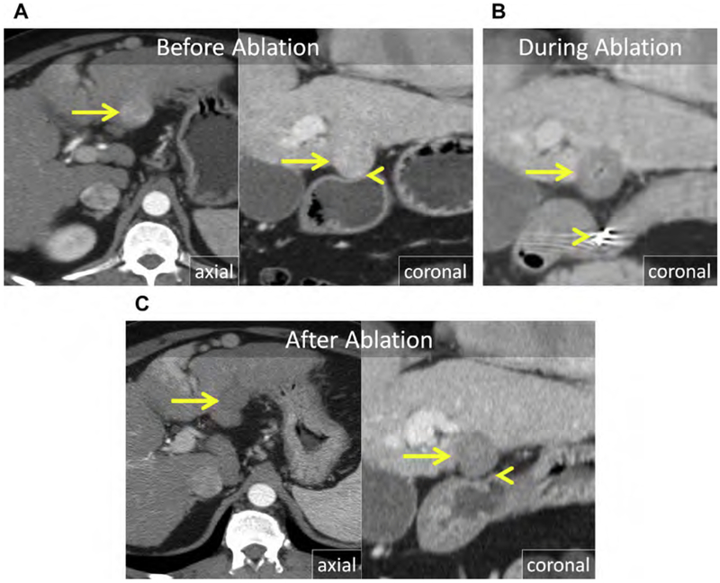
Fig. 21.
(A) Enhanced CT in the late arterial (axial) and portal venous phase (coronal reformat) shows an exophytic HCC projecting from the inferior left liver (arrows), abutting the gastric wall (arrowhead). (B) Coronal enhanced CT immediately following ablation. Two trocar needles (arrowhead) were used as a mechanical lever to displace the stomach from the index ablation (arrow). (C) Enhanced CT in the late arterial (axial) and portal venous phase (coronal reformat) in follow-up after MW ablation. There is no evidence for LTP (arrow) and the gastric wall is normal (arrowhead).

