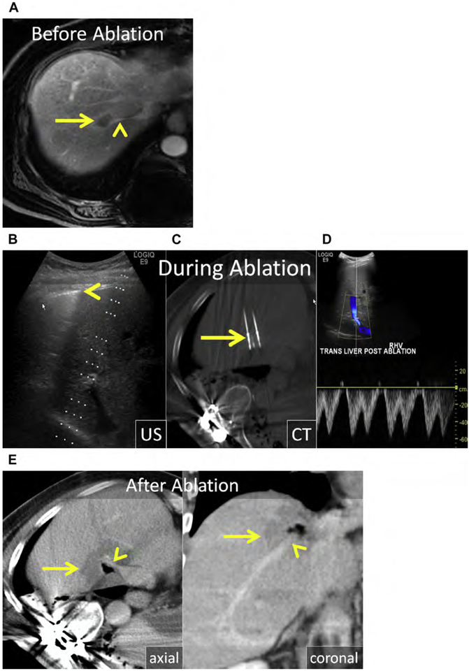
Fig. 4.
(A) CRLM in Couinaud segment VIII (arrow) abutting the right hepatic vein (arrowhead). (B, C) Three MW antennas were placed with US guidance into the mass and surrounding the right hepatic vein to overcome perfusion-mediated tissue cooling. (B) The edge of the lung (arrowhead) is easy to see and avoid during applicator placement. (C) Unenhanced CT was obtained to confirm precise antenna position (arrow). (D) Color and spectral Doppler was used intermittently during ablation to confirm patency of the right hepatic vein. (E) Axial and coronal enhanced CT immediately following MW ablation. The ablation encompasses the tumor and a margin of greater than 10 mm (arrow). The right hepatic vein remained patent (arrowhead).

